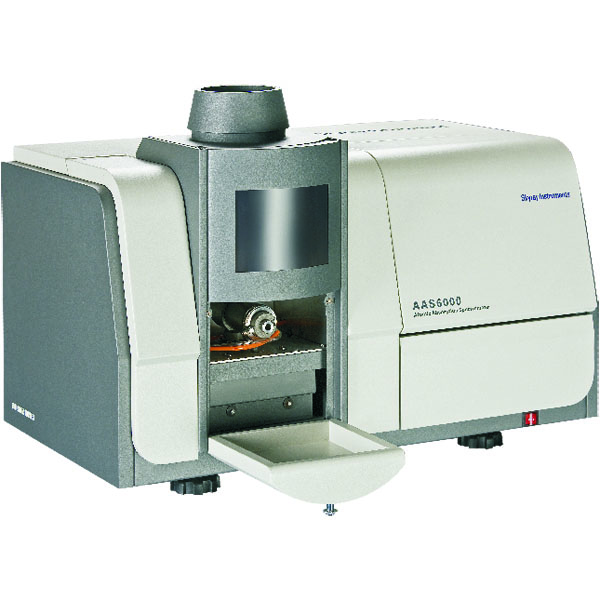
At the Very outset, it is important to comprehend the term spectrometer. A spectrometer is a device that divides and analyses the individual spectral components of a physical phenomenon to produce analytical results of curiosity. The spectrum — although most naturally related to mild by most — could also be mass, magnetic, electron etc. resulting in a huge range of kinds of spectrometry, such as optical spectrometry, photoelectron spectrometry, mass spectrometry etc. Optical spectrometry refers to the analysis of a light spectrum separated by wavelengths. It may be of two kinds — emission or absorption. An Atomic Optical/Emission Spectrometer AES / AAS is one that analyses an optical light spectrum emitted by an excited sample. The excitation could be by a range of ways, such as application of a spark, plasma, fire etc. Having said that, the expressional has become nearly ubiquitously employed by people to refer to this arc-spark AAS technique.

The key Fundamentals of spectrometry used in an arc/spark Optical Emission Spectrometer AAS are. Electrons in atoms absorb energy get excited and proceed into higher energy states also called orbits when energy is used. If this energy source is eliminated, the electrons fall in the ground state and release the absorbed energy in the form of photons. No two distinct element’s atoms can emit photons in exactly the exact same wavelength. Consequently, every wavelength is unique to one element alone. This means That once we know the wavelength of the photon emitted, we know which component is emitting it! In an Arc/spark AAS, the principles outlined above are leveraged to examine metallic by and large — but more on this later samples to evaluate precisely which elements exist in it and in what ratio. The output of this atomic absorption spectroscopy is a thorough evaluation of the elemental composition of the sample in weight percentages.
There is a need to spark the sample. The sample is therefore first ready, i.e., one face of the sample is made entirely uniform, clean, flat and as free of surface defects as possible. Suitable procedures of sample preparation has to be used for this. The prepared sample is then placed on the sample rack as shown below. The sample rack has a hole in it that the sample has to cover. Below this, there is an electrode at a predetermined distance in the sample’s exposed surface. This whole spark enclosure is filled with Argon when diagnosis is to be carried out. Then, a high current is applied to the sample. The Exceptionally high levels of DC current create a plasma from the Argon-purged air of the spark chamber, and a quick string of high-energy sparks is consequently created between the electrode and the sample. Application of those sparks causes a region of the sample to vaporise. The vaporised atoms in the plasma consume energy and their electrons move to greater energy-states with every spark. With each removal, the electrons go into ground state and emit photons.
